- News |
-
- Featured
-
Canada’s privacy commissioner launches investigation over the use..
As the years pass by, technology also widens, and more and more are being discovered. From simple gadgets..
- Business |
-
- Featured
-
Be an informer to I-T dept; earn up..
Sharing "specific information" with the income tax department about any benami..
- Tech & Industry |
-
- Featured
-
Gravitational wave event likely signaled birth of black..
The merger of two neutron stars that generated gravitational waves detected last year may have led to the birth..

- Entertainment |
-
- Topics
- Malayalam Film
- Media
- Music
- Youth
-
- Featured
-
Shawn Mendes Released Highly Anticipated Self-Titled Album Today
Los Angeles, CA : Multi-Platinum singer/songwriter Shawn Mendes released his highly anticipated self-titled third album today, via Island Records. Get..

-
- New Products |
-
- Topics
- General
-
- Featured
-
ZOTAC Introduces Its GeForce GTX 960 series graphics..
Dubai- ZOTAC International, a leading innovator and manufacturer of graphic cards and mini-

-
- Education |
-
- Topics
- Campus News
-
- Featured
-
ITM University, Gurgaon Student Palash Chhabra Represents Varsity..
New Delhi: Palash Chhabra, a student of ITM University,..

-
- Health |
-
- Topics
- Medical News
-
- Featured
-
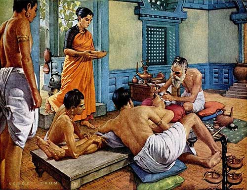
Maharshi Shushruta, The Great Grandfather of Surgery!
by Ayurvedacharya Dr.Hitesh Jani Dr.Hitesh Jani
-
- Tourism |
-
- Topics
- Travel
- Food&Beverages
- Hospitality
-
- Featured
-
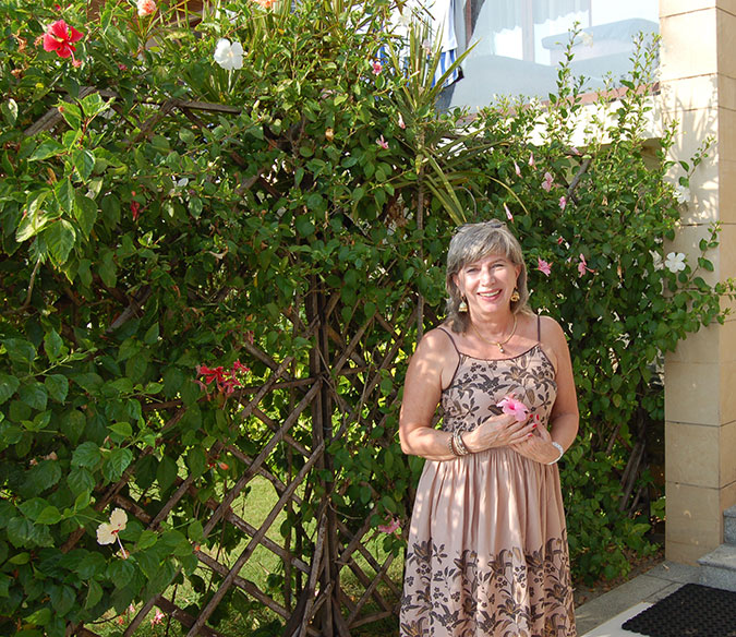
“Keraliya Ayurveda is Credible and Authentic”
Irina Gurjeva Irina Gurjeva is not just another vacationer in..
-
- Sports |
- Editor's column |
- Magazine |
Doctors at BLK Hospital Become World’s First to Remove Ruptured Hydatid Cyst using Cryoprobe
Published on September 17, 2020
NEW DELHI: In a first-ever recorded medical procedure of its kind, doctors at BLK Hospital removed a ruptured Hydatid cyst from the right lung of Ruhi-Un-Nisa, a 45-year-old woman from Srinagar, without conducting an open chest surgery. The cyst, formed by the larvae of tapeworms, was excised piecemeal by freezing it using a cryoprobe.
The patient showed initial symptoms and discomfort in July this year when she spit blood while coughing. She underwent a CT scan of the chest which revealed 43 X 35mm cyst (equivalent to a size of tennis ball) in superior segment of lower lobe of right lung. The patient followed the CT scan with a bronchoscopy procedure thereafter in Srinagar. However, her condition deteriorated, and she went into respiratory distress. Ruhi-Un-Nisa was unable to lie down and had not slept for close to two months. She had severe difficulty in breathing; she went to hospital in Kashmir where she was advised to rush to Delhi for emergency treatment.
The patient came with severe breathlessness and constantly felt a salty-bitter taste in her mouth. Doctors suspected the symptoms to be caused by a ruptured Hydatid cyst.
Hydatid cysts are known to trigger an extreme life-threatening allergic reaction in some patients. The patient immediately underwent bronchoscopy which revealed some whitish membranous structure inside her lung. This was one of a rare case of hydatid cyst whose removal was a challenge due to its positioning and breakage. These membranes are so fragile membrane that they tend to break easily on clasping.
The team led by Dr. Sandeep Nayar HOD, Chest & Respiratory Diseases, at BLK Hospital flushed out the spilled fluids/contents from both lungs. He then excised the ruptured membrane (of hydatid cyst) with the help of a Cryoprobe (a surgical probe to apply extreme cold to body tissues and freeze them). The membrane was frozen and extracted through the mouth and it gave immediate relief to the patient from her symptoms. Dr. Nayar performed this procedure through bronchoscope under local anesthesia. CT chest and bronchoscopy repeated after four days ensured that the lung was completely cleared of the cyst. The patient is now much better and almost without symptoms. She said this is the first time that she slept well after over two months!
Once ruptured, the contents of the hydatid cyst are released into the lungs and are usually coughed out. Hence, the salty taste was there in patient’s mouth. The spilled fluid can trigger anaphylactic shock in individuals that can lead to sudden death in as little as 6-8 hours. Luckily, the patient suffered no such shock even though the cyst had ruptured more than a month ago. All necessary precautions were taken during the procedure in case any such reaction occurs.
Said Dr. Sandeep Nayar HOD, Chest & Respiratory Diseases, at BLK Hospital: “Usually, the most common form of treatment of cystic echinococcosis is open surgical removal of the cyst, combined with chemotherapy. In this case, patient was very reluctant to undergo surgery and wanted immediate relief from her respiratory distress. After a detailed discussion with the CTVS team and family, we decided against surgery. Instead, we froze the ruptured cyst and removed it from the lung through the mouth without opening the chest. The entire procedure took around 45 minutes. If we had opted for surgery, we may have experienced complications, especially during these unprecedented times, so it was best avoided. This is the world’s first such procedure of its kind. The patient got instant relief after the procedure and will be on follow up de-worming medication for the next three months.”
He added that: “While hydatid cyst is not uncommon in India, it is routinely extracted surgically. While the patient clearly showed the symptoms of hydatid cyst, we did not know its present status and the damage it had caused. On investigation, we found that the hydatid had made a cavity in one of the lobes in the patient’s lungs. The cyst was punctured during previous bronchoscopy done in Srinagar and there was a constant fluid leak causing a constant bad taste in her mouth. This is when we decided to suck out the fluid and use the cryoprobe to freeze the residual cyst and its membranes and take it out through the patient’s mouth. This was done gradually over few attempts.”
Humans are accidental hosts of the long tapeworm and become infected by handling soil, dirt or animal hair that contains its eggs.
The highest occurrence of Hydatid cysts are found in states of Andhra Pradesh, Tamil Nadu and Jammu and Kashmir. These are caused due by larvae of a long tapeworm (Echinococcus granulosus) found in animals like dogs, sheep, cattle, goats, and pigs. The most common sites of occurrence inside human beings are the liver (55-70%) and lungs (18-35%). Unusual site can be kidneys, heart, spleen, brain, ovaries, etc (8-10%).
The eggs of the tapeworm contain an embryo which, when ingested by a human by eating unclean fruits and vegetables, contaminated by dog feces, develops into a hydatid cyst. This sac-like structure grows to 5 – 10 cm in size within the first year and can survive within human body for years, without showing any significant symptoms. These cysts sometimes become so large, by the end of several years or even decades they may contain several liters of fluid.